


Arthrosis of the hip joint is a degenerative-dystrophic pathology, characterized by destruction of hyaline cartilage.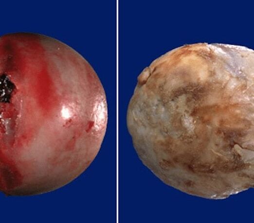 The disease develops gradually, accompanied by pain and decreased mobility. If there is no medical intervention in the early stages of arthrosis, after several years, femoral muscle atrophy occurs.
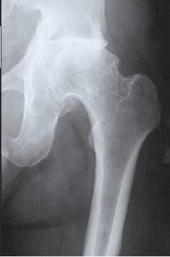 The injured limb is shortened, and fusion of the joint space causes immobilization of part or all of the hip joint. Pathological causes are previous injuries, curvature of the spine, systemic diseases of the musculoskeletal system.
Osteoarthritis is usually detected in middle -aged and elderly patients. Diagnosis is made based on the results of instrumental studies - radiography, MRI, CT, arthroscopy. Treatment of pathology of severity levels 1 and 2 is conservative. If ankylosis is detected or drug therapy is ineffective, surgical operations (arthrodesis, endoprosthetics) are performed.
Mechanisms of pathological development
The hip joint is formed by two bones - the ilium and the femur. The lower part of the ilium is represented by its body, which participates in articulation with the femur, forming the upper part of the acetabulum. During movement, the glenoid fossa does not move, and the femoral head moves freely. Such a device "hinges" the hip joint makes it possible to bend, bend, twist, induce abduction, addition of the hip. The fine, resilient hyaline cartilage, which lines the acetabulum and the femoral head provides an unobstructed sliding of the articular structure. Its main function is the redistribution of load during movement, the prevention of rapid wear of bone tissue.
Under the influence of external or internal factors, cartilage trophism is disrupted. It does not have its own circulatory system - synovial fluid supplies tissues with nutrients. With arthrosis, it thickens, becomes viscous. The lack of nutrients produced provokes drying of the hyaline cartilage surface. It is covered with cracks, which cause permanent tissue microtrauma during flexion or elongation of the hip joint. Cartilage becomes thinner and loses its protective properties. Defective bones to "adapt" to increased stress. And against the background of metabolic decline in tissues, destructive and degenerative changes develop.
Causes and provoking factors
Idiopathic or primary arthrosis develops for no reason. It is believed that the destruction of cartilage tissue occurs due to the natural aging of the body, slowing down the recovery process, decreased production of collagen and other compounds necessary for the regeneration of the structure of the hip joint. Secondary arthrosis occurs against the background of pathological conditions already present in the body. The most common causes of secondary illness include:
- previous injuries - damage to the ligament -tendon apparatus, rupture of muscles, their complete separation from the base of the bone, fractures, dislocations;
- violations of joint development, congenital dysplastic disorders;
- autoimmune pathology - rheumatoid, reactive, psoriatic arthritis, systemic lupus erythematosus;
- non -specific inflammatory diseases such as purulent arthritis;
- specific infections - gonorrhea, syphilis, brucellosis, ureaplasmosis, trichomoniasis, tuberculosis, osteomyelitis, encephalitis;
- endocrine system dysfunction;
- degenerative -dystrophic pathology - osteochondropathy of the femoral head, osteochondritis dissecans;
- joint hypermobility, due to the production of "super-extensible" collagen, provokes their excessive movement, ligament weakness.
Since the cause of the development of arthrosis can be hemarthrosis (bleeding in the cavity of the hip joint), provoking factors include disorders of hematopoiesis. Prerequisites for the onset of the disease are overweight, excessive physical activity, inactive lifestyle. Its development is due to improper organization of sports training, lack of dietary foods with high content of micro -elements, vitamins and soluble in water. Postoperative arthrosis occurs several years after surgery, especially if it is accompanied by the cutting of a large amount of tissue. Hyaline cartilage trophism is frustrated with frequent hypothermia, living in an unpleasant environment, and working with toxic substances.
Arthrosis of the hip joint cannot be inherited. But in the presence of certain congenital features (metabolic disorders, skeletal structure), the likelihood of its development increases significantly.
Symptoms
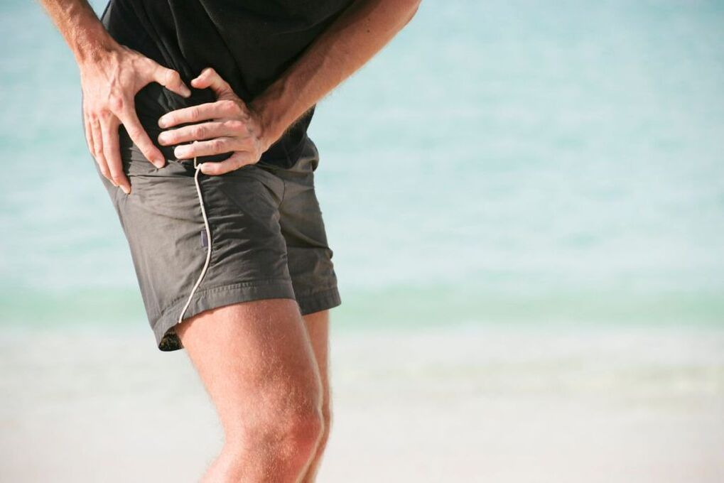
The main symptom of arthrosis of the hip joint is pain when walking in the hip area, radiating to the groin, knee joint. A person experiences stiffness of movement, stiffness, especially in the morning. To stabilize the joints, the patient begins to drown, his gait changes. Over time, due to muscle atrophy and articulatory deformities, the limb feels short. Another pathological hallmark is the limitation of hip abduction. For example, difficulties arise when trying to sit on a bench with legs apart.
For arthrosis of first severity, periodic pain occurs after intense physical exercise. They are localized in the articulation area and disappear after a long rest.
With second -degree arthrosis of the hip joint, the severity of the pain syndrome increases. Discomfort occurs even at rest, extending to the thighs and groin, increasing with weight lifting or increased motor activity. To relieve pain in the hip joint, a person begins to drown almost invisibly. Limitation of movement in the joints was observed, especially during abduction and internal rotation of the thigh.
Third -degree arthrosis is characterized by persistent severe pain that does not subside day and night. Difficulties arise when moving, therefore, when walking, one is forced to use a cane or crutches. The hip joints are stiff, there is significant atrophy of the muscles of the back, thighs, and legs. Due to the weakness of the abductor’s femoral muscle, the pelvic bone does not move in the frontal area. To compensate for the shortening of the leg, the patient leans on the injured limb while moving. This causes a strong shift in the center of gravity and an increase in pressure on the joints. At this stage of arthrosis, significant joint ankylosis develops.
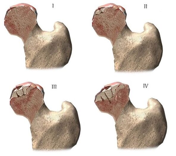
| Degrees | Radiographic signs |
| The first | The change is insignificant. The joint gap is moderately, unevenly narrowed, there is no destruction of the femoral surface. Small bone growth is observed on the outer edge or in the acetabulum |
| The second | The height of the joint space is significantly reduced due to its uneven fusion. The head of the femur bone is moved upwards, deformed, enlarged, its contour becomes uneven. Bone growth is formed on the inner and outer edge surfaces of the glenoid fossa |
| Third | There is a combination of whole or partial joint space. The femoral head is strongly raised. Much of the bone growth is located on all surfaces of the acetabulum |
Diagnostics
When making a diagnosis, the doctor takes into account the clinical manifestations of pathology, anamnesis, the results of external examination of the patient and instrumental studies. Radiography is the most informative. With its help, the condition of the hip joint is assessed, the degree of its travel, the degree of damage to the cartilage tissue, and in some cases the cause of its development can be known. If the cervical-diphyseal node is enlarged, and the acetabulum oblique and flat, then with a high degree of probability, dysplastic congenital changes in the articulation are likely to occur. Perthes disease or juvenile epiphysiolysis is indicated by a disturbed shape of the hip bone. Radiography can reveal post-traumatic arthrosis, although there is no previous trauma in the anamnesis. Other diagnostic methods are also used:
- CT helps detect the growth of bone plate edges, osteophytes are formed;
- MRI is performed to assess the state of connective tissue structures and the degree of their involvement in pathological processes.
If necessary, the inner surface of the joint is examined with an arthroscopic instrument. Differential diagnosis is performed to exclude gonarthrosis, lumbosacral or thoracic osteochondrosis. Pain in arthrosis can be disguised as a clinical manifestation of radicular syndrome caused by nerve traps or inflammation. It is often possible to exclude neurogenic pathology with the help of a series of tests. Arthrosis of the hip joint is necessarily distinguished from trochanteric bursitis of the hip joint, ankylosing spondylitis, reactive arthritis. To exclude autoimmune pathology, biochemical studies of blood and synovial fluid were performed.
Drug treatment tactics
Medical treatment aims to improve the well -being of patients. For this, drugs from various clinical and pharmacological groups are used:
- nonsteroidal anti -inflammatory drugs (NSAIDs) - Nimesulide, Ketoprofen, Diclofenac, Ibuprofen, Meloxicam, Indomethacin, Ketorolac. To relieve acute pain, injectable solutions are used, and pills, pills, ointments, gels help relieve severe or moderate pain;
- glucocorticosteroids - Triamcinolone, Dexamethasone, Hydrocortisone. They are used in the form of intra-articular blockade in combination with the anesthetic Procaine, Lidocaine;
- muscle relaxation - Baclofen, Tizanidine. They are included in the treatment regimen for skeletal muscle spasms, pinching of sensitive nerve endings;
- drugs that increase blood circulation in the joints - nicotinic acid, Aminophylline, Pentoxifylline. Is prescribed to patients to improve tissue trophism, prevent the progression of the disease;
- chondroprotectors. Effective only on stage 1 and 2 arthrosis.
Rubbing the ointment with a warming effect helps relieve mild pain. The active ingredients of external agents are capsaicin, cinquefoil, camphor, menthol. This substance is characterized by irritating, annoying, local analgesic effect. Pressing on the joints with Dimethylsulfoxide, medical bile will help relieve the swelling, swelling of the thighs in the morning. Patients are recommended classical massage, acupressure or vacuum for coxarthrosis. Daily exercise therapy is a better prevention of the development of arthrosis.
Surgical intervention
With the effectiveness of conservative therapy or a complicated pathological diagnosis by ankylosis, surgery is performed. It is impossible to restore cartilage tissue in joints damaged by arthrosis without prosthetic surgery, but with the right treatment approach, adhering to all medical prescriptions, maintaining a proper lifestyle, doing therapeutic exercises, regular massage courses, taking vitamins and proper nutrition, youcan stop the process of lesions and destruction of cartilage and hip joints.